Knee
Knee Anatomy
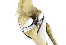
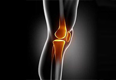
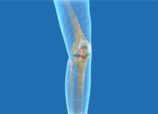
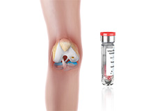
The knee is a complex joint made up of different structures - bones, tendons, ligaments, and muscles. They all work together to maintain the knee’s normal function and provide stability to the knee during movement.
Having a well-functioning healthy knee is essential for our mobility and ability to participate in various activities. Understanding the anatomy of the knee enhances your ability to discuss and choose the right treatment procedure for knee problems with your doctor.
Bones of the Knee
The knee is a hinge joint made up of two bones, the thighbone (femur) and shinbone (tibia). There are two round knobs at the end of the femur called femoral condyles that articulate with the flat surface of the tibia called the tibial plateau. The tibial plateau on the inside of the leg is called the medial tibial plateau and on the outside of the leg, the lateral tibial plateau.
The two femoral condyles form a groove on the front (anterior) side of the knee called the patellofemoral groove. A small bone called the patella sits in this groove and forms the kneecap. It acts as a shield and protects the knee joint from direct trauma.
A fourth bone called the fibula is the other bone of the lower leg. This forms a small joint with the tibia. This joint has very little movement and is not considered a part of the main joint of the knee.
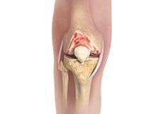
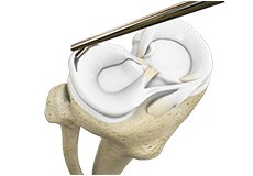
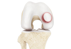
Articular Cartilage and Menisci of the Knee
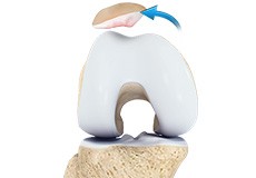
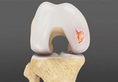
Movement of the bones causes friction between the articulating surfaces. To reduce this friction, all articulating surfaces involved in the movement are covered with a white, shiny, slippery layer called articular cartilage. The articulating surface of the femoral condyles, tibial plateaus and the back of the patella are covered with this cartilage. The cartilage provides a smooth surface that facilitates easy movement.
To further reduce friction between the articulating surfaces of the bones, the knee joint is lined by a synovial membrane that produces a thick clear fluid called synovial fluid. This fluid lubricates and nourishes the cartilage and bones inside the joint capsule.
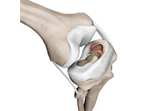
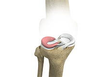
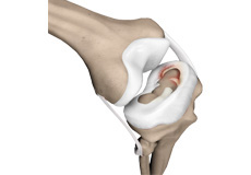
Within the knee joint, between the femur and tibia, are two C-shaped cartilaginous structures called menisci. Menisci function to provide stability to the knee by spreading the weight of the upper body across the whole surface of the tibial plateau. The menisci help in load-bearing i.e. it prevents the weight from concentrating onto a small area, which could damage the articular cartilage. The menisci also act as a cushion between the femur and tibia by absorbing the shock produced by activities such as walking, running and jumping.
Ligaments of the Knee
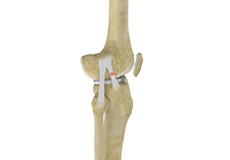
Ligaments are tough bands of tissue that connect one bone to another bone. The ligaments of the knee stabilize the knee joint. There are two important groups of ligaments that hold the bones of the knee joint together, collateral and cruciate ligaments.
Collateral ligaments are present on either side of the knee. They prevent the knee from moving too far during side to side motion. The collateral ligament on the inside is called the medial collateral ligament (MCL) and the collateral ligament on the outside is called the lateral collateral ligament (LCL).
Cruciate ligaments, present inside the knee joint, control the back-and-forth motion of the knee. The cruciate ligament in the front of the knee is called anterior cruciate ligament (ACL) and the cruciate ligament in the back of the knee is called posterior cruciate ligament (PCL).
Muscles of the Knee
There are two major muscles in the knee - the quadriceps and the hamstrings, which enable movement of the knee joint. The quadriceps muscles are located in front of the thigh. When the quadriceps muscles contract, the knee straightens. The hamstrings are located at the back of the thigh. When the hamstring muscles contract, the knee bends.
Tendons of the Knee
A tendon is a tissue that attaches a muscle to a bone. The quadriceps muscles of the knee meet just above the patella and attach to it through a tendon called the quadriceps tendon. The patella further attaches to the tibia through a tendon called the patella tendon. The quadriceps muscle, quadriceps tendon, and patellar tendon all work together to straighten the knee. Similarly, the hamstring muscles at the back of the leg are attached to the knee joint with the hamstring tendon.
Conditions
Knee Fracture
A fracture is a condition in which there is a break in the continuity of the bone. In younger individuals, these fractures are caused by high energy injuries, as from a motor vehicle accident. In older people, the most common cause is a weak and fragile bone.
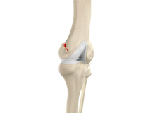
Patella Fracture
The kneecap or patella forms a part of the knee joint. It is present at the front of the knee, protecting the knee and providing attachment to various muscle groups of the thigh and leg. The undersurface of the kneecap and the lower end of the femur are coated with articular cartilage, which helps in smooth movement of the knee joint. A fracture in the kneecap is rare but common in adult males.
Pediatric ACL Tears
The anterior cruciate ligament (ACL) is a ligament that provides stability, reduces stress and prevents the knee from rotating or slipping out of position while jumping, running and landing. This ligament can tear during sports activities and exercise, as a result of a non-contact twisting injury, and is becoming a common injury in children.
Knee Injury
Pain, swelling, and stiffness are the common symptoms of any damage or injury to the knee. If care is not taken during the initial phases of injury, it may lead to joint damage, which may end up destroying your knee.
Knee Sprain
Knee sprain is a common injury that occurs from overstretching of the ligaments that support the knee joint. A knee sprain occurs when the knee ligaments are twisted or turned beyond its normal range, causing the ligaments to tear.
Knee Infection
Knee infection is a serious medical condition that needs immediate treatment. Infection may occur followed by a knee replacement surgery or trauma and is usually caused by bacteria. Infection may spread to the space of the knee joint or deep layers of your knee causing serious complications.
ACL Tears
The anterior cruciate ligament (ACL) is one of the major ligaments of the knee. It is located in the middle of the knee and runs from the femur (thighbone) to the tibia (shinbone). The ACL prevents the tibia from sliding out in front of the femur. Together with the posterior cruciate ligament (PCL), it provides rotational stability to the knee.
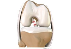
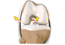
Meniscal Injuries
The knee is one of the most complex and largest joints in the body and is very susceptible to injury. The meniscus is a small, C-shaped piece of cartilage in the knee. Each knee consists of two menisci, medial meniscus on the inner aspect of the knee and the lateral meniscus on the outer aspect of the knee. The medial and lateral menisci act as a cushion between the thighbone (femur) and shinbone (tibia).

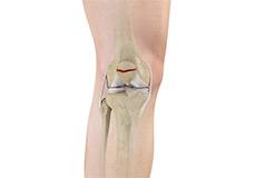
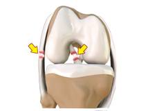
Meniscal Tears
The two wedge-shaped cartilage pieces present between the thighbone and the shinbone are called meniscus. They stabilize the knee joint and act as shock absorbers. What is a Meniscal Tear?
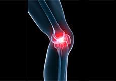
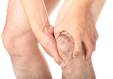
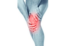
Knee Pain
Knee pain is a common condition affecting individuals of various age groups. It not only affects movement but also impacts your quality of life. An injury or disease of the knee joint or any structure surrounding the knee can result in knee pain. A precise diagnosis of the underlying cause is important to develop an appropriate treatment plan.
Multiligament Instability
The knee is a complex joint of the body that is vital for movement. The four major ligaments of the knee are anterior cruciate ligament (ACL), posterior cruciate ligament (PCL), medial collateral ligament (MCL) and lateral collateral ligament (LCL). They play an important role in maintaining the stability of the knee. A multiligament injury is a tear in one or more ligaments of the knee, which affects the knee stability.
Knee Arthritis
The joint surface is covered by a smooth articular surface that allows pain-free movement in the joint. Arthritis is a general term covering numerous conditions where the joint surface or cartilage wears out. This surface can wear out for several reasons; often the definite cause is not known. Arthritis often affects the knee joint.
PCL Injuries
Posterior cruciate ligament (PCL), one of the four major ligaments of the knee, is situated at the back of the knee. It connects the thighbone (femur) to the shinbone (tibia). The PCL limits the backward motion of the shinbone.
Patellar Instability
The patella is a small piece of bone in front of the knee that slides up and down the groove in the femur bone during bending and stretching movements. The ligaments on the inner and outer sides of the patella hold it in the femoral groove and avoid dislocation of the patella from the groove.
Lateral Meniscus Syndrome
The knee joint is formed by the union of two bones, namely the femur (thighbone) and the tibia (lower leg bone). At the junction of these two bones is a cartilage called meniscus, which acts as a shock absorber. There are two menisci – the lateral and medial menisci. The lateral meniscus is the outer meniscus of the knee joint and gives a cushioning effect during weight-bearing activities.
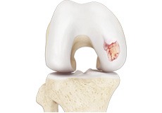
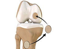
Articular Cartilage Injury
Articular or hyaline cartilage is the tissue lining the surface of the two bones in the knee joint. Cartilage helps the bones move smoothly against each other and can withstand the weight of the body during activities such as running and jumping. Articular cartilage does not have a direct blood supply to it so has little capacity to repair itself.
Knee Osteoarthritis
Osteoarthritis also called degenerative joint disease, is the most common form of arthritis. It occurs most often in older people. This disease affects the tissue covering the ends of bones in a joint (cartilage).In a person with osteoarthritis, the cartilage becomes damaged and worn out causing pain, swelling, stiffness and restricted movement in the affected joint.
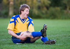
Knee Sports Injuries
Trauma is any injury caused during physical activity, motor vehicle accidents, electric shock, or other activities. Sports trauma or sports injuries refer to injuries caused while playing indoor or outdoor sports and exercising. Sports trauma can result from accidents, inadequate training, improper use of protective devices, or insufficient stretching or warm-up exercises. The most common sports injuries are sprains and strains, fractures, and dislocations.
Multiligament Knee Injuries
Injury to more than one knee ligament is called a multiligament knee injury and may occur during sports or other physical activities.
Anterior Knee Pain
The knee joint is a large, complex joint in the body that comprises of three bones, i.e. the lower end of the thighbone or femur, the upper end of the shinbone or tibia, and the kneecap or patella. The patella moves over the joint and allows bending of the knee and straightening of the leg.
Recurrent Patella Dislocation
The patella (kneecap) is a small bone that shields your knee joint. It is present in front of your knee, on a groove called the trochlear groove that sits at the junction of the femur (thighbone) and tibia (shinbone). Articular cartilage presents below the patella and end of the femur cushion and helps the bones glide smoothly over each other when the legs move.
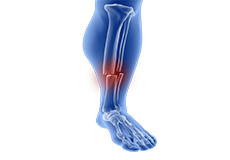
Tibia Fractures
A crack or break in the tibia is referred to as a tibial fracture. The tibia is the most frequently fractured long bone of the body. It normally takes a great amount of force for a fracture of the tibia to occur.
Procedures
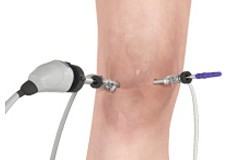
Knee Arthroscopy
Knee arthroscopy is a common surgical procedure performed using an arthroscope, a viewing instrument, to diagnose or treat a knee problem. It is a relatively safe procedure and you will usually be discharged from the hospital on the same day of surgery.
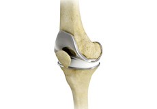
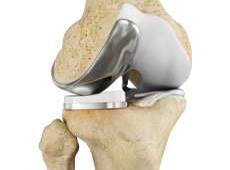
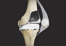
Total Knee Replacement
The knee is made up of the femur (thighbone), tibia (shinbone) and patella (kneecap). The two menisci, soft cartilage between the femur and tibia, serve as a cushion and help absorb shock during motion. Arthritis (inflammation of the joints), injury or other diseases of the joint can damage this protective layer, causing extreme pain and difficulty in performing daily activities.
Outpatient Total Knee Replacement
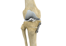
Total knee replacement is the surgical treatment for knee arthritis, where the damaged knee is removed and replaced with an artificial knee implant. Traditionally performed as an inpatient procedure, total knee replacement surgery is now being conducted on an outpatient basis, allowing you to go home on the same day of the surgery.
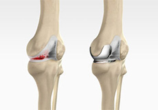
Minimally Invasive Knee Joint Replacement
Total knee replacement is a very successful surgical treatment for knee arthritis. Over the years, minimally invasive knee replacement surgical techniques have been developed to lessen tissue trauma and improve patient outcomes. This minimally invasive approach involves much smaller incisions than the usual 10-12 inch incisions used in the traditional knee replacement and spares the quadriceps muscle and tendon, which control bending of the knee, from being cut to access the knee joint.
Arthroscopic Debridement
Osteoarthritis is a most common form of arthritis which affects the articular cartilage (tissue covering the ends of the bones) of the knee and also other joints such as shoulder, hip, ankle, and foot. The articular cartilage cushions the joint so that there is smooth and pain-free movement between the bones in the joint.
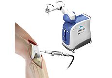
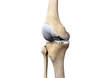
Robotic Assisted Knee Replacement
The knee is made up of the femur (thighbone), the tibia (shinbone) and patella (kneecap). The two menisci, the soft cartilage between the femur and tibia, serve as a cushion and helps absorb shock during motion. Arthritis (inflammation of the joints), injury or other diseases of the joint can damage this protective layer of cartilage, causing extreme pain and difficulty in performing daily activities.
Unicompartmental/Partial Knee Replacement
Arthritis is the inflammation of a joint that causes pain, swelling (inflammation) and stiffness.
Osteoarthritis is the most common form of knee arthritis, in which the joint cartilage gradually wears away. It most often affects older people. In a normal joint, articular cartilage allows for smooth movement within the joint, whereas in an arthritic knee the cartilage itself becomes thinner or completely absent.
Unicondylar knee Replacement
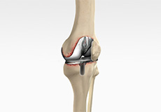
Unicompartmental knee replacement is a minimally invasive surgery in which only the damaged compartment of the knee is replaced with an implant. It is also called a partial knee replacement. The knee can be divided into three compartments: patellofemoral, the compartment in front of the knee between the knee cap and thigh bone, medial compartment, on the inside portion of the knee, and lateral compartment which is the area on the outside portion of the knee joint.

Outpatient Unicondylar Knee Replacement
A unicondylar knee replacement, also known as unicompartmental or partial knee replacement, is a procedure to replace a portion of the damaged knee joint with a prosthetic implant to relieve pain and improve function of the knee joint. Traditionally performed as an inpatient procedure, advances in technology have allowed this procedure to be performed in a minimally invasive manner on an outpatient basis allowing patients to go home the same day of the surgery
Partial Medial Knee Replacement
The knee is made up of the femur (thighbone), the tibia (shinbone), and patella (kneecap). The meniscus, the soft cartilage between the femur and tibia, serves as a cushion and helps absorb shock during motion. The knee can be divided into three compartments:
Partial Knee Resurfacing
Partial knee replacement is an alternative to total knee replacement in patients with arthritis on only one side of the knee. Partial knee replacement is a surgical procedure which involves resurfacing and replacement of only the diseased surface of the joint instead of the entire joint.
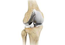
Tricompartmental Knee Replacement
The knee is made up of the femur (thighbone), the tibia (shinbone), and patella (kneecap). The meniscus, the soft cartilage between the femur and tibia, serves as a cushion and helps absorb shock during motion. The knee can be divided into three compartments:
Revision Knee Replacement
The knee joints are lined by soft articular cartilage that cushion the joints and aid in smooth movement of the joint bones. Degeneration of the cartilage due to wear and tear leads to arthritis, which is characterized by severe pain.
Meniscal Surgery
A meniscus tear is the commonest knee injury in athletes, especially those involved in contact sports. A sudden bend or twist in your knee can cause the meniscus to tear. This is a traumatic meniscal tear. The elderly are more prone to degenerative meniscal tears as the cartilage wears out and weakens with age.
A torn meniscus causes pain, swelling, stiffness, catching or locking sensation in your knee, making you unable to move your knee through its complete range of motion.
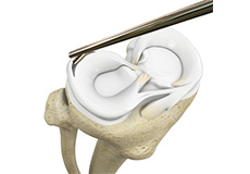
Meniscectomy
Meniscectomy is a surgical procedure indicated in individuals with torn meniscus where the conservative treatments are a failure to relieve the pain and other symptoms. Meniscectomy is recommended based on the ability of meniscus to heal, patient’s age, health status, and activity level.
Partial Meniscectomy
Partial meniscectomy is a surgical procedure to remove the torn portion of the meniscus from the knee joint.
Meniscal tears can occur at any age, but are more common in athletes playing contact sports. These tears are usually caused by twisting motion or over-flexing of the knee joint.
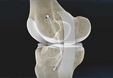
PCL Reconstruction
The posterior cruciate ligament (PCL), one of four major ligaments of the knee, is situated at the back of the knee. It connects the thighbone (femur) to the shinbone (tibia). The PCL limits the backward motion of the shinbone.
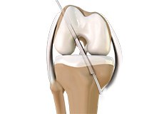
ACL Reconstruction
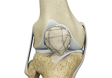
The anterior cruciate ligament (ACL) is one of the major stabilizing ligaments in the knee. It is a strong rope-like structure located in the center of the knee, running from the femur to the tibia. When this ligament tears, unfortunately, it does not heal on its own, and often leads to the feeling of instability in the knee.
ACL Reconstruction of Patellar Tendon
The anterior cruciate ligament is one of the four major ligaments of the knee that connects the femur (thighbone) to the tibia (shinbone) and helps stabilize the knee joint. The anterior cruciate ligament prevents excessive forward movement of the lower leg bone (tibia) in relation to the thighbone (femur) as well as limits rotational movements of the knee.
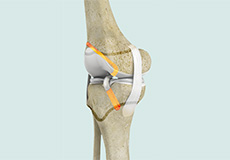
MCL Reconstruction
An injury to the MCL may occur as a result of direct impact to the knee. An MCL injury can result in a minor stretch (sprain), or a partial or complete tear of the ligament.
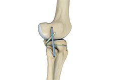
ORIF of the Knee Fracture
A knee fracture is a break in the continuity of bone within the knee. This can involve the tibia (shinbone), kneecap (patella), or femur (thigh bone).
ORIF refers to open reduction and internal fixation. It is a surgical procedure employed for the treatment of a fracture not amenable to non-surgical conservative treatment.
Knee Osteotomy
Knee osteotomy is a surgical procedure in which the upper shinbone (tibia) or lower thighbone (femur) is cut and realigned. It is usually performed in arthritic conditions affecting only one side of your knee. The aim is to take pressure off the damaged area and shift it to the other side of your knee with healthy cartilage.
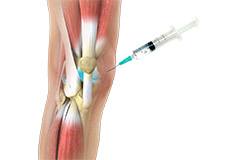
Viscosupplementation
Viscosupplementation refers to the injection of a hyaluronan preparation into the joint. Hyaluronan is a natural substance present in the joint fluid that assists in lubrication. It allows the smooth movement of the cartilage-covered articulating surfaces of the joint.
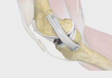
Meniscal Transplantation
Meniscal transplantation is a surgical procedure to replace the damaged meniscus of the knee with healthy cartilage. The meniscus is a C-shaped cartilage ring that acts as a cushion between the shinbone and the thighbone.
Chondroplasty
Chondroplasty is a surgical procedure to repair and reshape damaged cartilage in a joint. The procedure involves smoothing degenerative cartilage and trimming any unstable flaps of cartilage.
Matrix Induced Autologous Chondrocyte Implantation
Matrix-Induced autologous chondrocyte implantation is an innovative, FDA-approved cartilage restoration procedure that uses your own cells to repair cartilage defects in your knee. It can alleviate knee pain, help you regain function and may even delay or prevent arthritis.
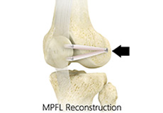
Medial Patellofemoral Ligament Reconstruction
Medial patellofemoral ligament reconstruction is a surgical procedure indicated for severe patellar instability.
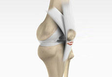
Posterolateral Corner Reconstruction
Posterolateral corner injury is damage or injury to the structures of the posterolateral corner. The structures of the posterolateral corner include the lateral collateral ligament, the popliteus tendon, and the popliteo-fibular ligament. Injuries to the posterolateral corner most often occur with athletic trauma, motor-vehicle accidents, and falls.
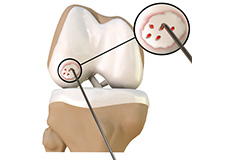
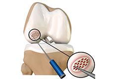
Cartilage Microfracture
Cartilage microfracture is a surgical procedure performed to replace the worn-out articular cartilage with new cartilage. It is usually performed to treat small areas of cartilage damage usually caused by sports or traumatic injuries. It is not indicated if you have advanced arthritis of the knee.
Osteochondral Allografts
Osteochondral grafting is a method of treating cartilage injuries that expose underlying bone. An osteochondral allograft is a piece of tissue containing bone and cartilage that is taken from a deceased donor to replace damaged cartilage that lines the ends of bones in a joint.
Others
Nonoperative Treatments for ACL Injuries
The ACL (anterior cruciate ligament) is one of the four major ligaments located within the knee joint. It connects the femur (thighbone) to the tibia (shinbone). It plays a key role in holding the two bones within the knee and keeping the joint stable while your knee moves back and forth.